LOVE AND DEATH AT THE NIH

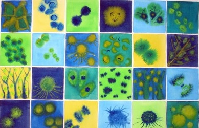
I first started experimenting with watercolor about 10 years ago, and from the beginning got into “wet in wet technique.” To paint “wet in wet” you paint a base color and then add other colors to it while it’s still wet. This allows the different colors to bleed into each other, making interesting patterns. People who saw my wet-in-wet work at shows kept mentioning how much it looked like cells under a microscope, so I found some images of cells in mitosis, or cell division, and discovered that they did indeed look a lot like what I was doing. After looking at the images I began actually trying to paint cells, but I guess I’ve been painting them for about a decade.
Last winter, when DC was buried under several feet of snow, I decided to finally make a move into online art sales, opening up a shop on Makers Market, a juried online marketplace with a scientific bent. The cell pieces were instantly my most popular, so I’ve been making more and more cell images. I’ve been showing my work, mainly at art festivals around DC, for about 10 years. Selling online connected me to a whole new audience and provided a creative shot in the arm. A bunch of biologists bought my work, and some of them suggested new subjects, like bacteria or blood cells. One buyer pointed me toward “brainbows” – a series of images of mouse brain cells dyed in bright colors. I loved them, and the images inspired me to begin painting brain cells.
I was talking about this new work with an artist friend, Sean Hennessey, who mentioned that he was having a show of his work at the National Institutes of Health (NIH) and suggested that my cell paintings would be a good fit. The curator at NIH agreed, so I started to conceptualize this show. I decided that a whole show of mitosis paintings would be a little boring, and I wanted the exhibit to have a stronger theme.

I got to thinking about how activity at the cellular level underlies major upheavals in our lives, like falling in love, giving birth and dying. I decided to divide my paintings into two groups – love and death. It’s a kind of ridiculously grandiose theme, but I’m a lover of opera and Russian literature, and heck, go big or go home, right?
I learned a whole lot of anatomy and biology while painting these pictures. I looked up microscopic photographs in books and on the web and did many practice paintings, trying to get the balance right between accuracy and artistry. The “love” paintings focus on the cells that are involved in attraction and desire – the skin, eyes, and ears, the brain and the circulatory system. I’m very proud of my blood vessels, especially my abdominal aorta. (That’s not technically a cell, but ok, too bad.)
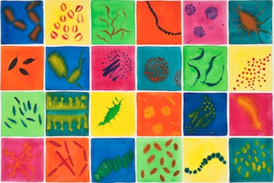
For the “death” paintings, I depicted three microscopic killers – bacteria, viruses and cancer. The cancer piece really hit home, because I lost both my parents to pancreatic cancer over the last decade, and I had never really approached it in my art before. And I included two mitosis paintings, because cell division underlies the whole process of life and death.
I’m really happy with the work I’ve put together for this exhibit, and I feel like it’s opened new doors for me creatively. The exhibit opens January 14th and will be up through March 5, 2011 at the NIH Clinical Center West Gallery (10 Center Drive, Bethesda MD 20892.) For more info about getting to NIH, see:http://www.nih.gov/about/visitor/index.htm
(Originally published in BourgeonOnline)